

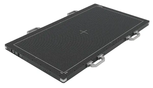
The device incorporates an active area of 430 × 860 mm, equivalent to twice the image area of a conventional 43 × 43 cm detector, allowing for expanded anatomical coverage in a single acquisition. This new model expands Detection Technology's X-Panel XL family into the field of medical imaging and combines a large-format TFT sensor with a cesium iodide (CsI) scintillator and high-speed readout electronics.
The X-Panel 4386a FPM is designed to image larger anatomical areas without the need for multiple acquisitions.
Depending on the system's geometry, its larger surface area can allow for an expanded reconstructed field of view from approximately 25 cm to 50 cm, compared to a conventional 43 × 43 cm detector. This capability can help reduce truncation artifacts and improve the efficiency of CBCT imaging procedures.
Its intended applications include whole-body and longitudinal axis CBCT, intraoperative and mobile CBCT systems, image-guided radiotherapy, and weight-bearing studies.
Amorphous silicon sensor and CsI scintillator
The detector uses an amorphous silicon TFT sensor fabricated on a single sheet of glass, combined with a directly deposited CsI scintillator. This architecture eliminates junctions and gaps within the active imaging area, reducing the possibility of junction-related artifacts and providing a uniform surface for calibration.
The device also incorporates a mounting system for an anti-scatter grid (ASG), designed to reduce scattered radiation in CBCT applications with wide fields of view. According to Tuomas Holma, product manager at Detection Technology, the combination of an extra-large TFT sensor, a CsI scintillator, and fast-readout electronics provides medical equipment manufacturers with greater flexibility in developing new imaging systems.
High-speed dynamic acquisition and dual-sided reading
The X-Panel 4386a FPM incorporates high-speed readout electronics and a dual-detector readout system, enabling dynamic acquisition across its large active area. It also supports pixel binning and region of interest (ROI) selection modes, allowing manufacturers to tailor spatial resolution and acquisition speed to the specific requirements of each clinical protocol.
The ability to perform shorter acquisitions can help reduce image degradation caused by movement and improve patient comfort.
Two versions for CBCT and image-guided radiotherapy
Detection Technology offers two configurations of the new detector, adapted to different integration needs:
X-Panel 4386a FPM-CSV-TG: optimized for CBCT medical tomography applications.
X-Panel 4386a FPM-CSV-TG-S: incorporates internal shielding to protect the electronics from scattered megavoltage (MV) radiation in radiotherapy environments.
The second version is designed to obtain kilovoltage (kV) X-ray CBCT images in image-guided radiotherapy procedures, including patient positioning and adaptive radiotherapy workflows.
Both configurations share the following specifications:
Active area: 430 × 860 mm.
Pixel size: 140 µm.
Image matrix: 3,072 × 6,144 pixels.
Sensor technology: Amorphous silicon TFT on single glass.
Scintillator: cesium iodide (CsI).
Data interface: 10G Ethernet.
Development compatibility: SDK for Windows and Linux.
Integration into next-generation medical equipment
The new detector is geared towards original equipment manufacturers (OEMs) developing medical imaging systems with higher requirements for anatomical coverage and dynamic acquisition. The combination of an extra-large active surface, fast readout, and different acquisition modes allows the device to be adapted to various CBCT and radiotherapy architectures.
With the X-Panel 4386a FPM, Detection Technology expands its catalog of X-ray detectors for medical applications, offering a solution for large field of view CBCT systems, intraoperative imaging and image-guided radiotherapy.