

Thanks to augmented reality, the doctor will be able to see, through a pair of glasses, information about the device, including the exact location for the puncture, its depth and angle, the patient's respiratory cycle, and, simultaneously, access to data and graphs on the patient's vital signs and other relevant parameters. The new system will also allow for early biopsies of very small tumors, where current systems are ineffective, thus enabling treatment of the disease in its initial stages.
The prototype will be able to use images from different medical modalities, such as computed tomography (CT), magnetic resonance imaging (MRI), and positron emission tomography (PET), etc. This will represent a significant leap forward compared to current biopsies due to its greater precision and, importantly, will allow doctors to act sooner, without having to wait for a tumor to grow before performing a biopsy. All of this will be achieved using commercially available technology.
Evolved version
The new system is an evolution of a first biopsy guidance device in cancer patients using positron emission imaging (PET), developed by the same team and which last summer was awarded at the Massachusetts Institute of Technology within the framework of an International Mentoring Program, in which it participated thanks to the sponsorship of the Foundation for Innovation and Foresight in Health in Spain, FIPSE.
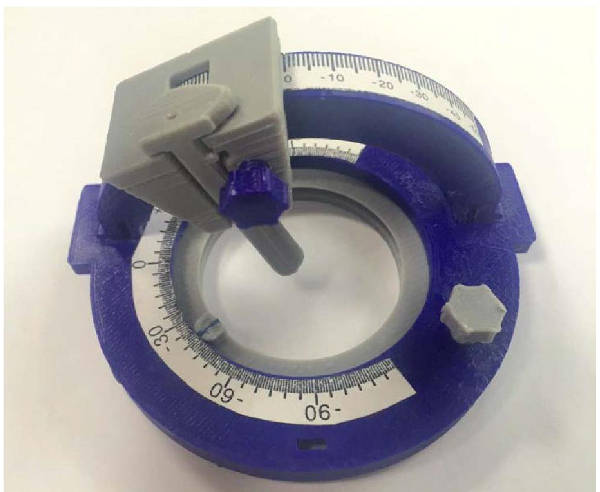
The initial, pre-commercial system consists of a stereotactic frame, which, together with software, virtualizes the position of the biopsy needle, enabling highly accurate cell or tissue extraction from the patient. The stereotactic frame parameters the patient's space using a spherical coordinate system, with precise housing for positron-emitting sources and an adhesive base for positioning and securing it to the anatomical area to be biopsied. The biopsy needle is graduated with a metric scale and contains a specific housing for the precise positioning of a positron-emitting source. The multiplatform software allows for obtaining the precise spherical coordinates of the target area for biopsy and verifying needle positioning from positron emission tomography (PET) images.
It is useful in patients who do not present with a structural anatomical radiological alteration that would guide the biopsy; and in patients with radiological structural lesions but with heterogeneous positron emission tomography (PET) scans. It allows obtaining a sample from an area of higher metabolic activity, avoiding punctures of necrotic or hemorrhagic tissue that are not useful for histological diagnosis.
The use of this device will increase the precision and accuracy of biopsies in cancer patients. It prevents non-diagnostic biopsies and reduces procedure time. Patient safety is enhanced by avoiding repeat biopsies and minimizing the radiation dose received from CT scans. As a result, the cost-effectiveness of image-guided biopsy procedures will improve.
The device is highly useful for guiding biopsies with conventional X-ray imaging, since by replacing the positron-emitting elements with radiopaque elements, the kit can be adapted for guiding the biopsy needle using CT imaging, where the number of cases is significantly higher. In both situations, PET-guided and CT-guided punctures, the aim is to increase the accuracy and precision of the punctures, avoiding repeat procedures and their adverse effects.
The team
The research team at the University Hospital of Araba consists of Ignacio Tobalina Larrea, specialist in Nuclear Medicine and Internal Medicine, Head of the Nuclear Medicine Service of the Araba Integrated Health Organization; María Ángeles García Fidalgo, specialist in Hospital Radiophysics and Head of the Medical Physics and Radiological Protection Service of the Araba Integrated Health Organization; Gaspar Sánchez Merino, specialist in Hospital Radiophysics; and Jesús Cortés Rodicio, specialist in Hospital Radiophysics.